NPL - Ultrasound Breast Density measurement
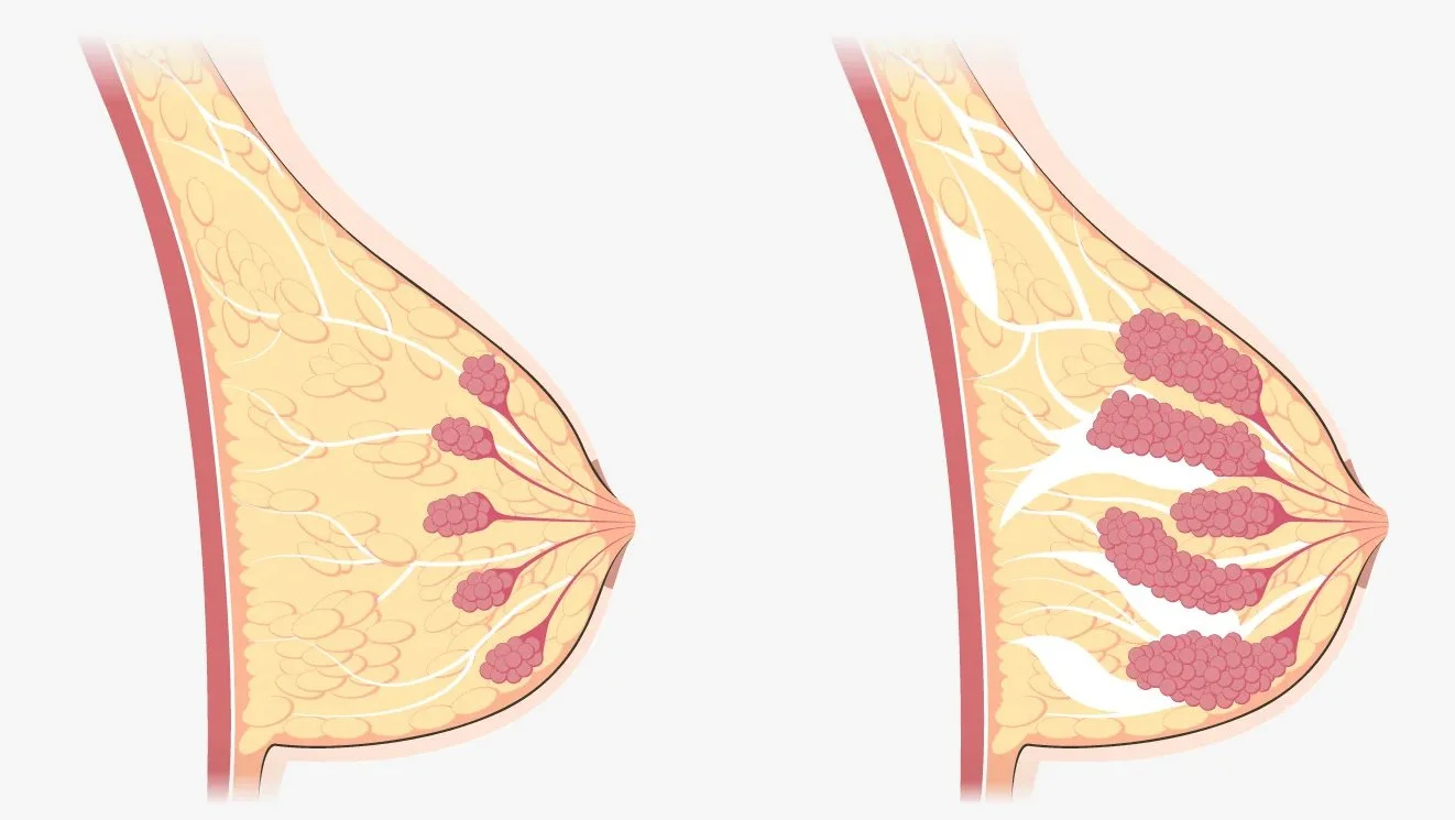
Breast density describes the relative ratio of fibroglandular tissue to fat in breasts. Whilst it can be expressed as a simple percentage, many women don’t know what theirs is.
Women with “very dense breasts are up to four times more likely to develop breast cancer than women with low breast density” [1] so it’s really important they are better informed, and can then access the diagnostic interventions they might need.
Researchers at NPL proposed a novel technological approach to density measurement and came to Designworks to support implementing it for undertaking volunteer participant research. Their long-term aim is providing a more accessible method of density measurement at point of care, without the pain and radiation dose inherent in mammography.
Being informed at a younger age could help those women with higher breast density know that they are at higher risk, and would benefit from earlier and more frequent examination.
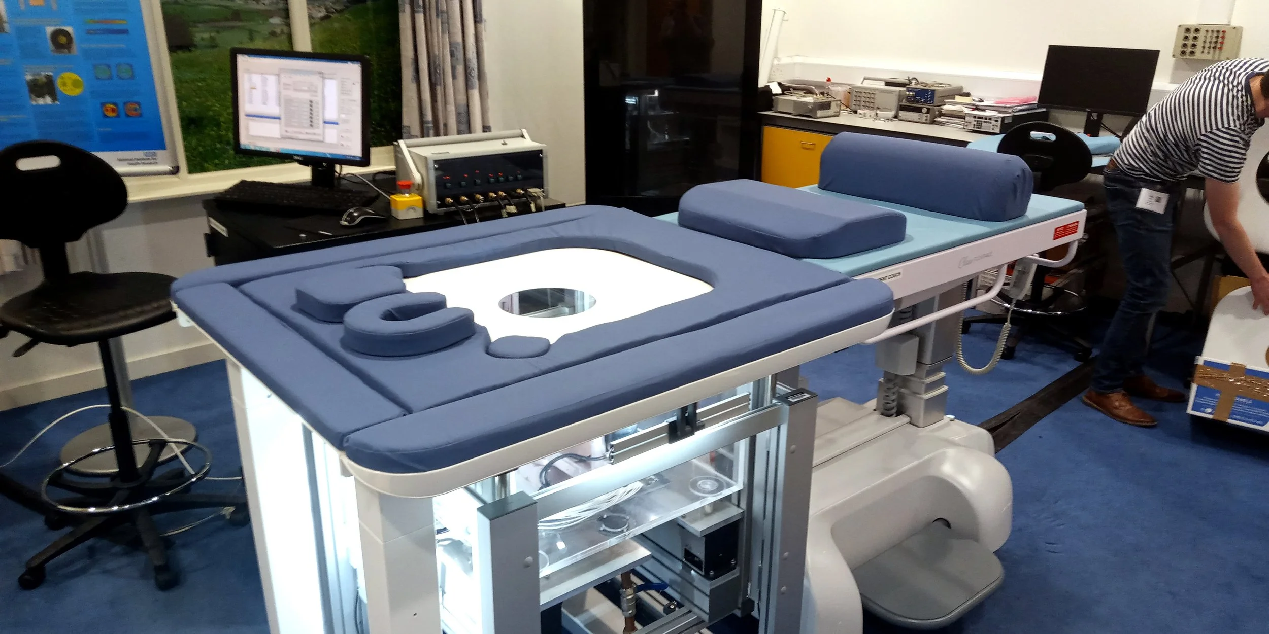
Designworks had supported NPL’s ultrasound and underwater acoustics department in the past with their PiUCT breast imaging platform. For that development, we provided a modular suite of accessories to improve the comfort of participants using the research platform, as well as a design concept with a full-scale looks-like model.
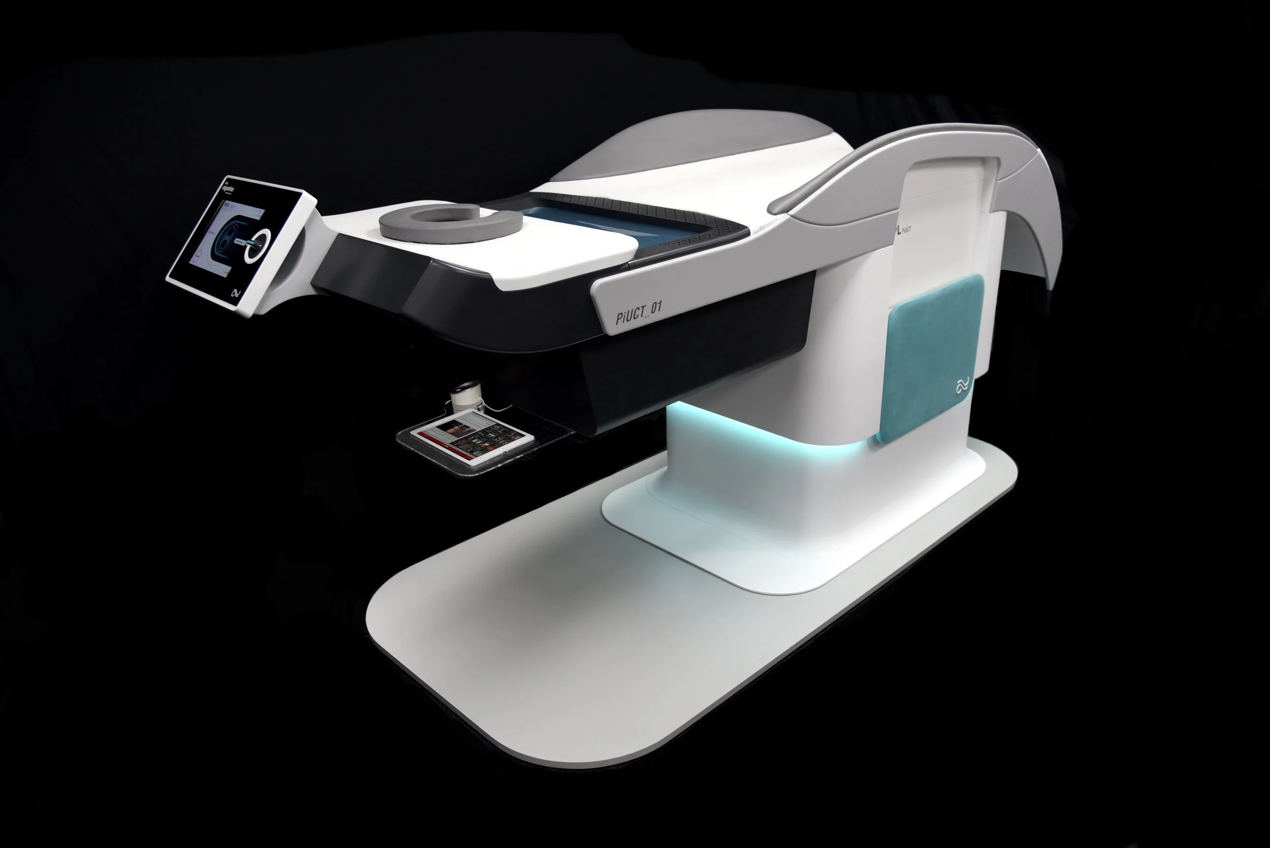
For this next requirement to measure breast density, we aimed to deliver these both simultaneously; a practical, functioning research platform that also demonstrated an initial pass at how a commercial device might be realised.
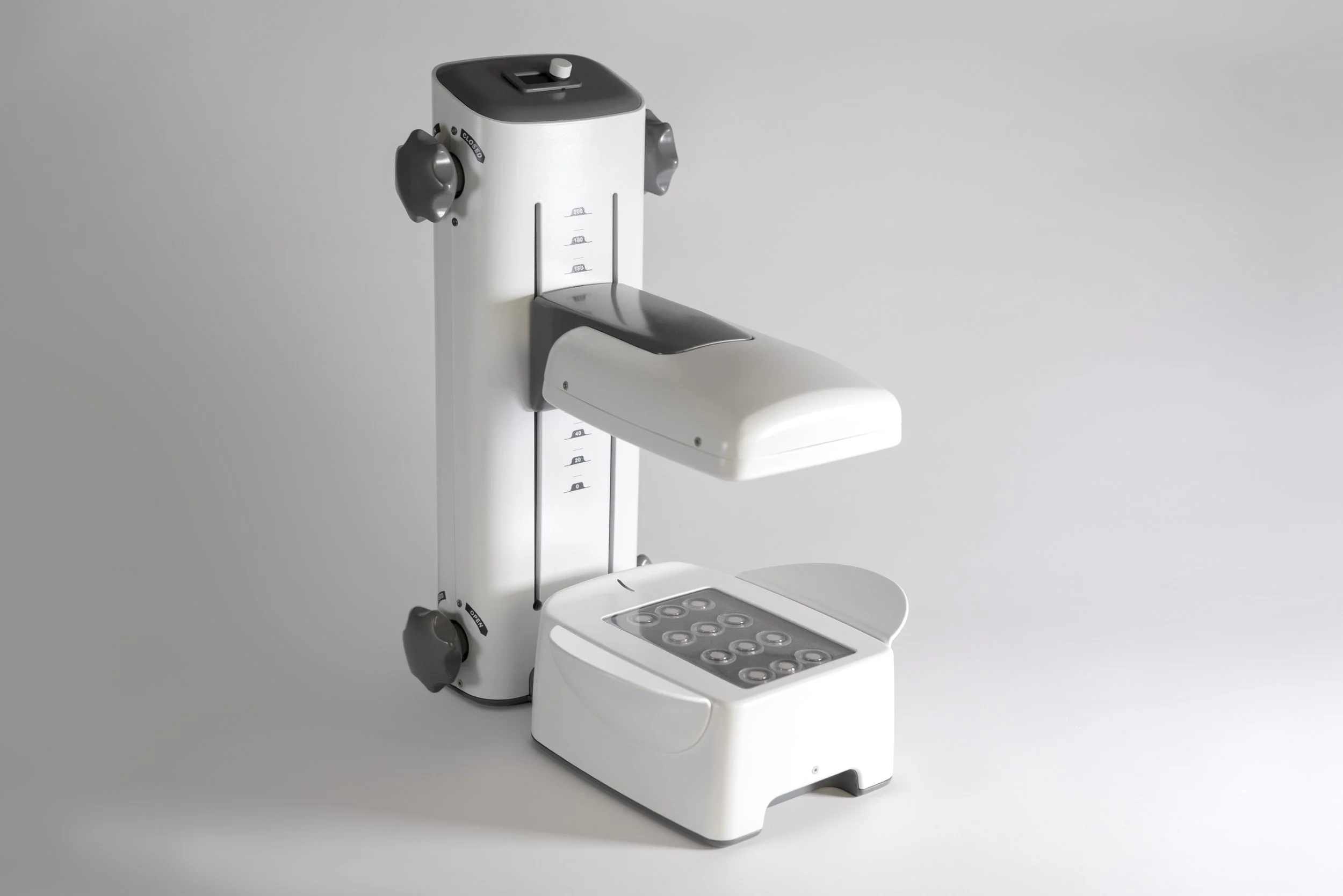
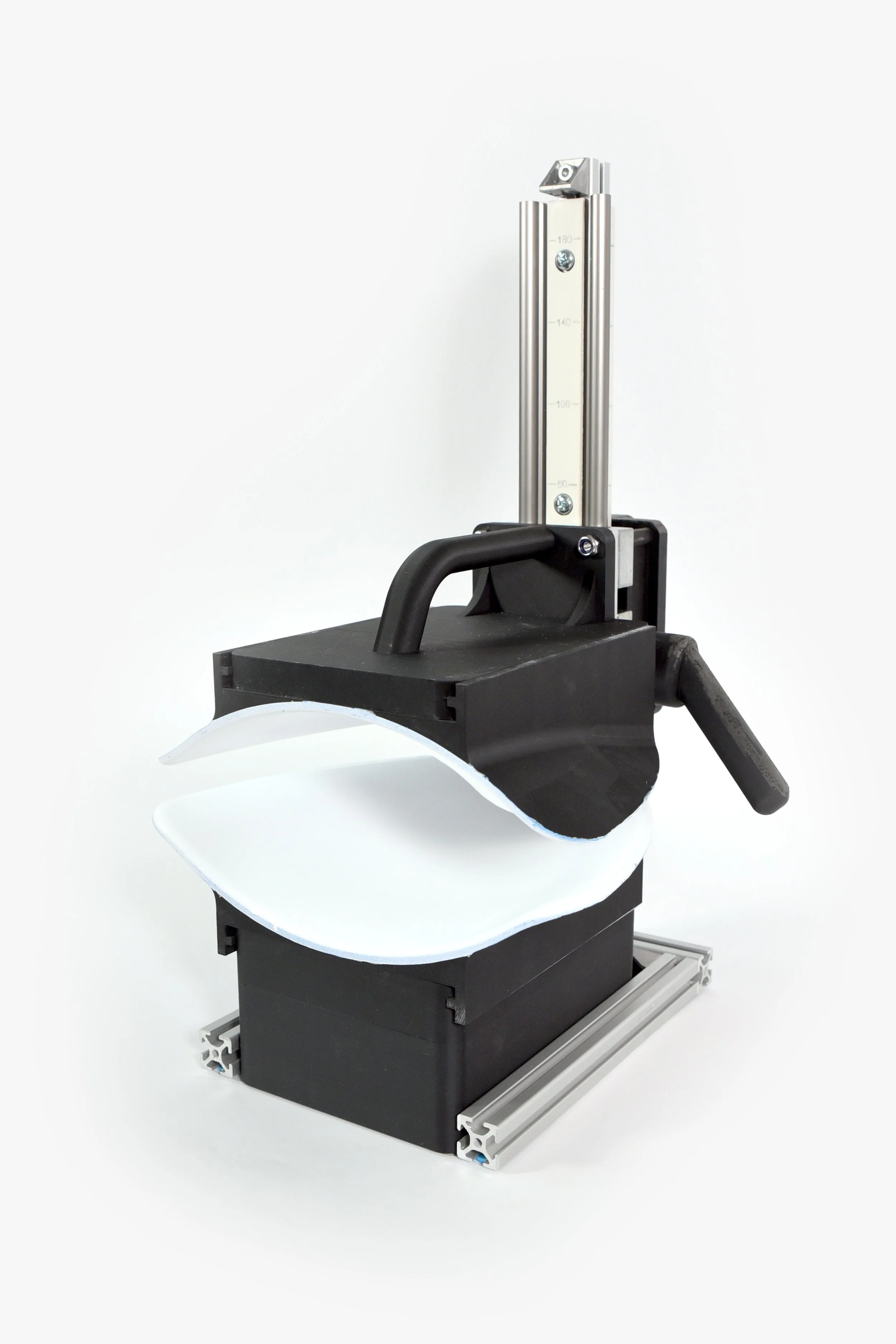
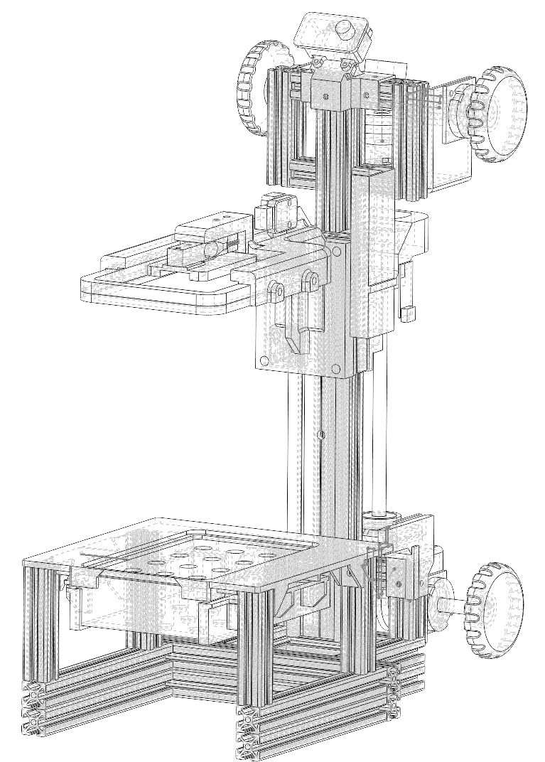
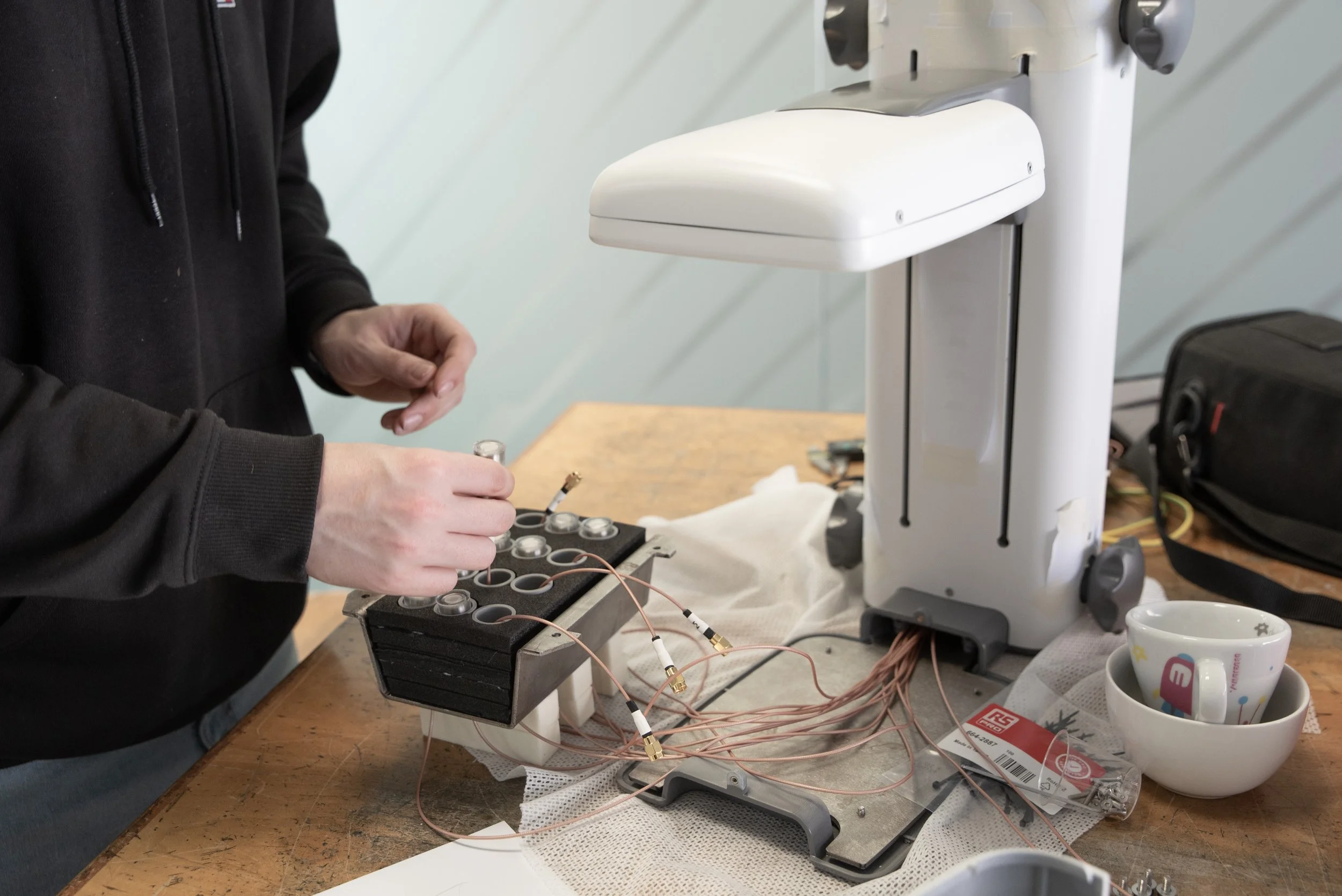
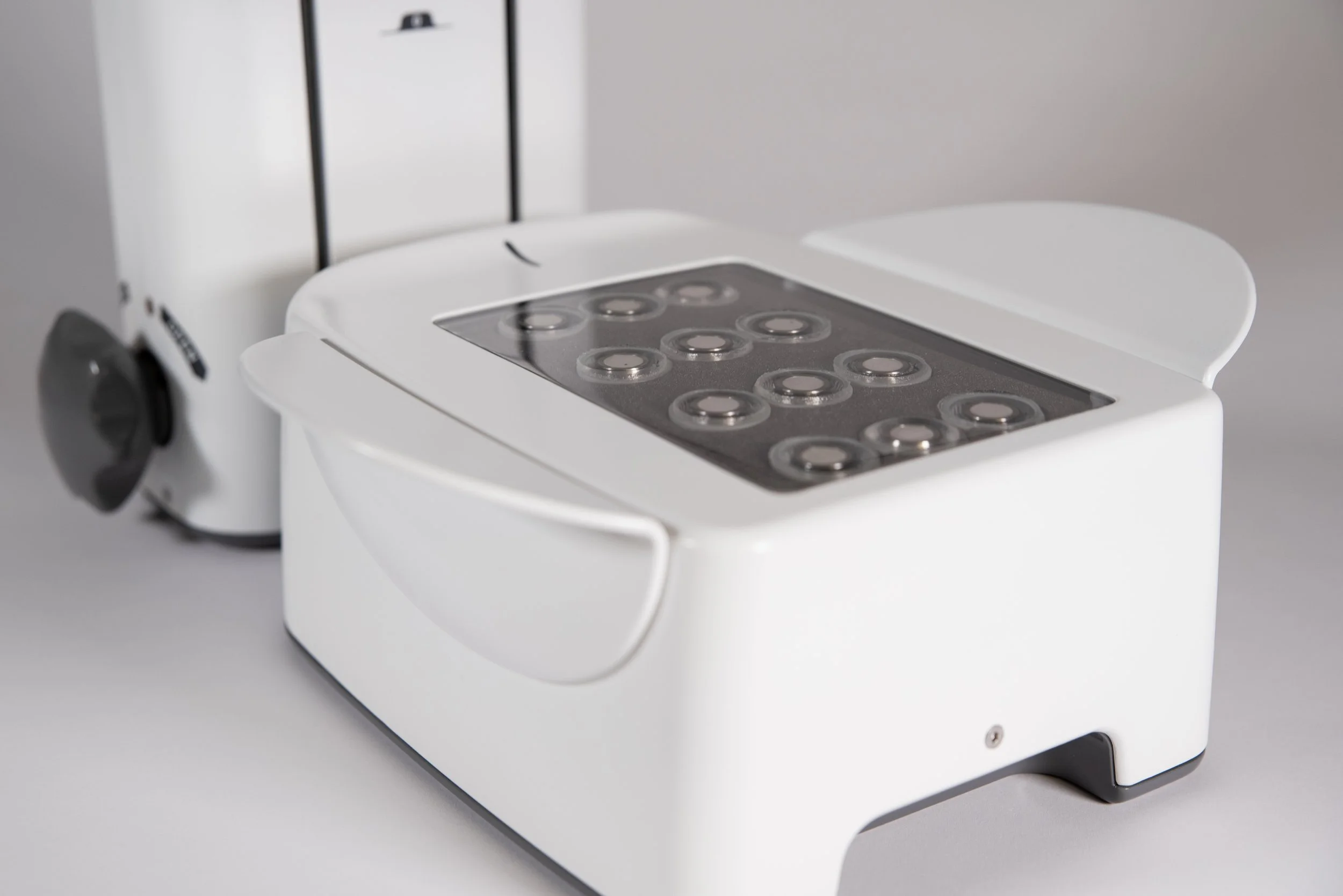
NPL specified an ultrasound transducer (emitter) to be included in an array of twelve and procured a custom-made sensor, but required a physical platform to use them.
To take a suitable reading, the sensor and array of transducers must close around the breast, expelling all air in-between.
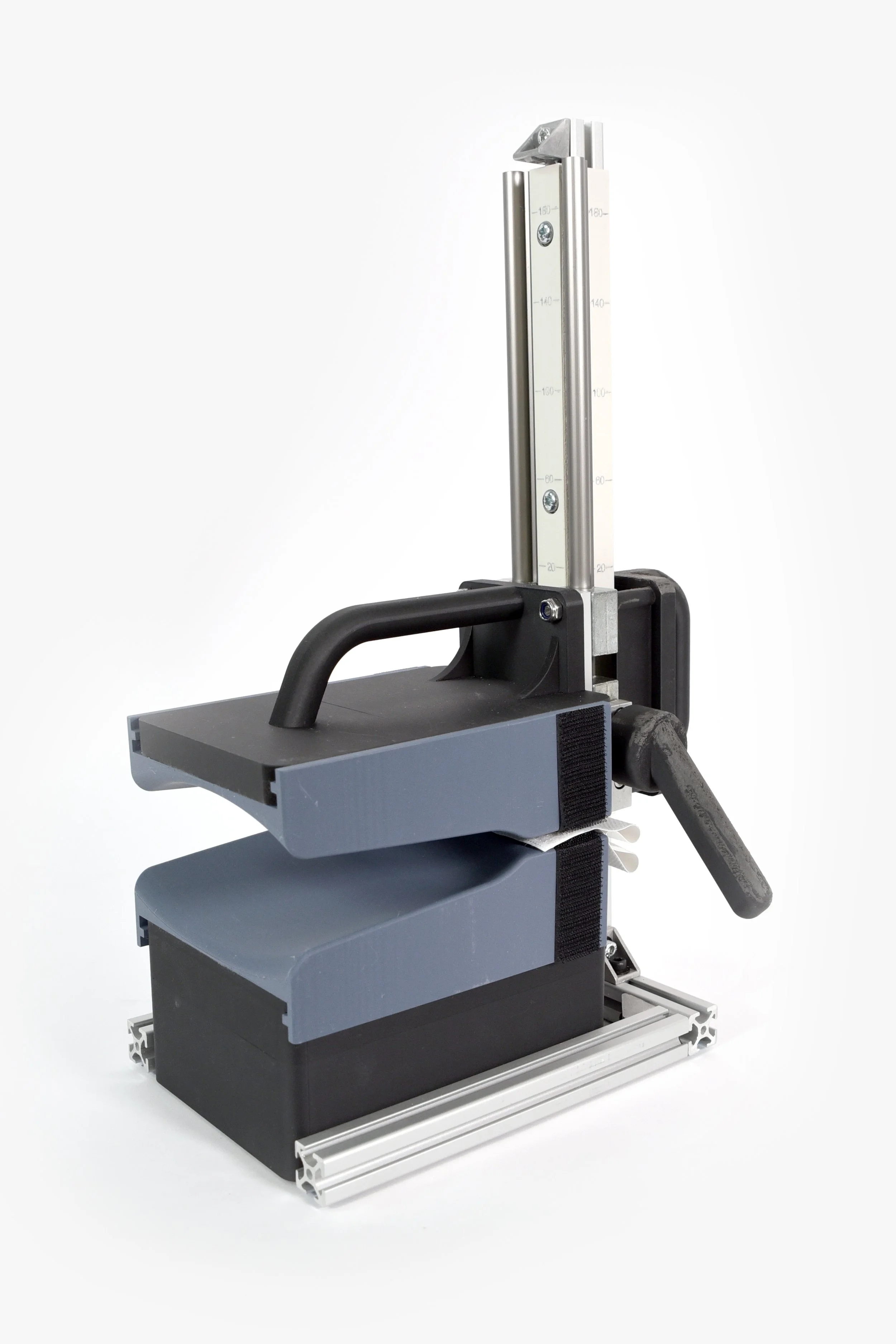
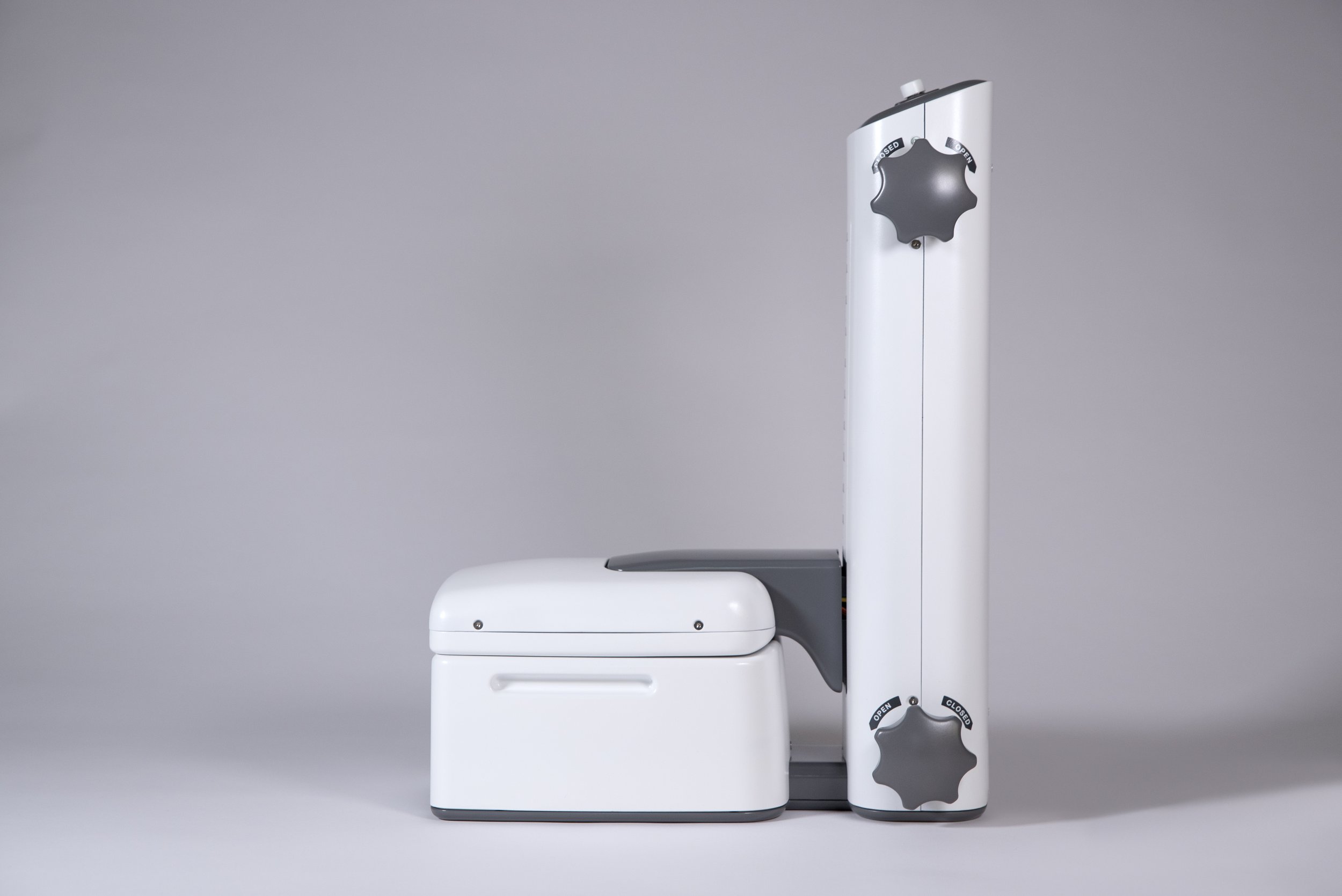
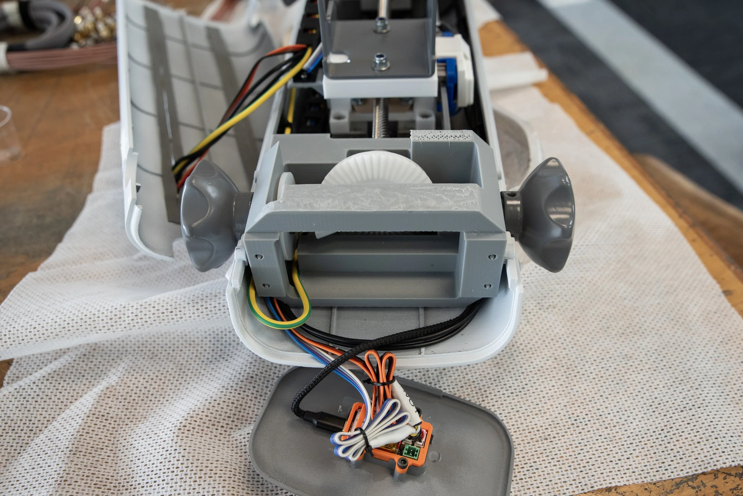
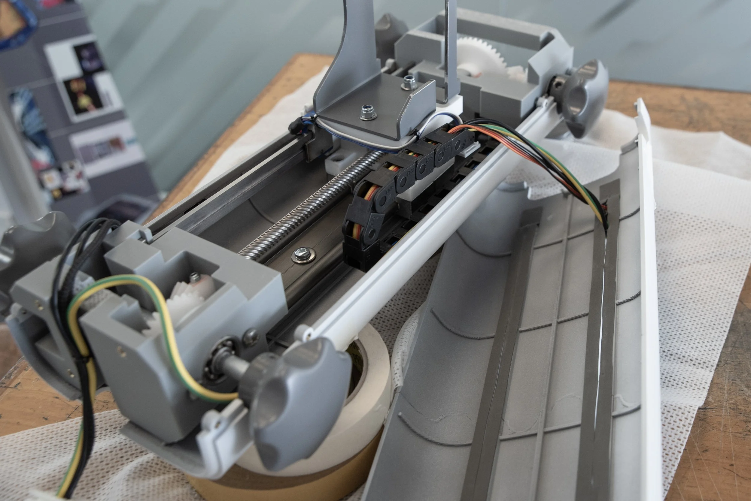
As the transducer array would be more densely wired, we elected to keep it static and move the sensor linearly to meet it, testing a range of configurations with low-fidelity modular rigs, then early visibility prototypes.
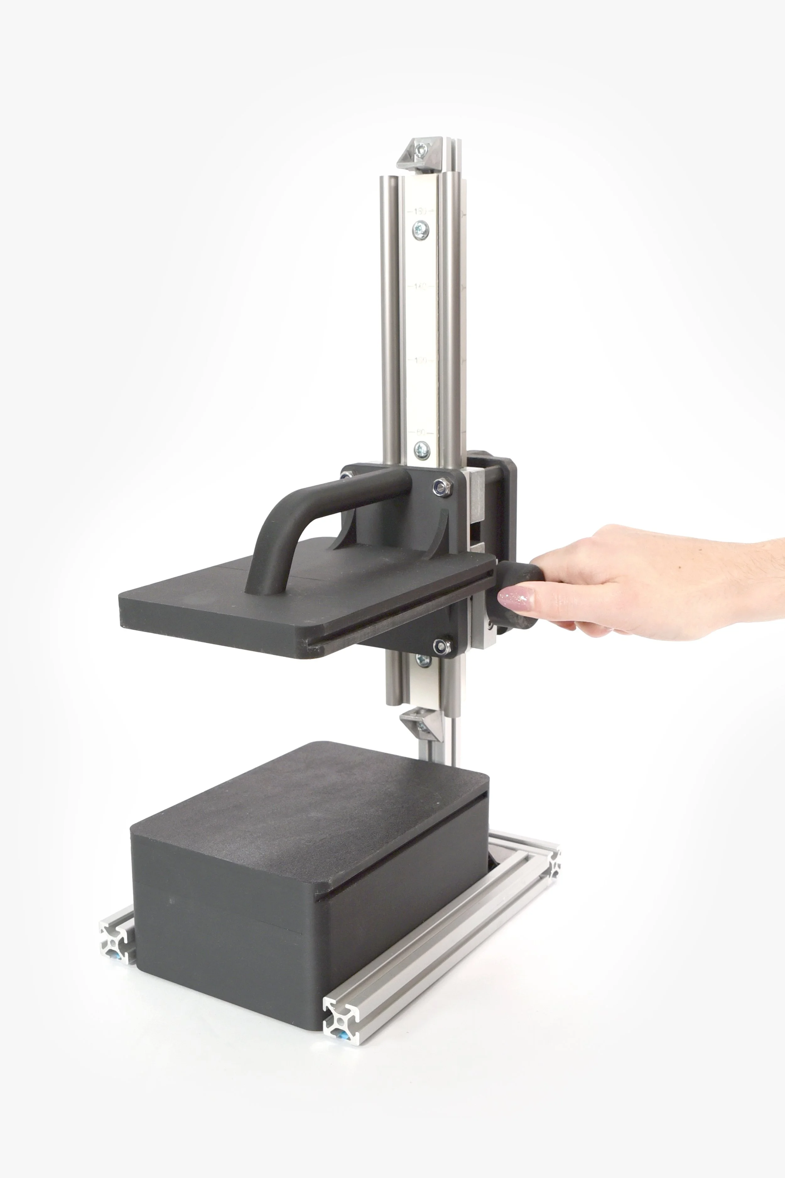
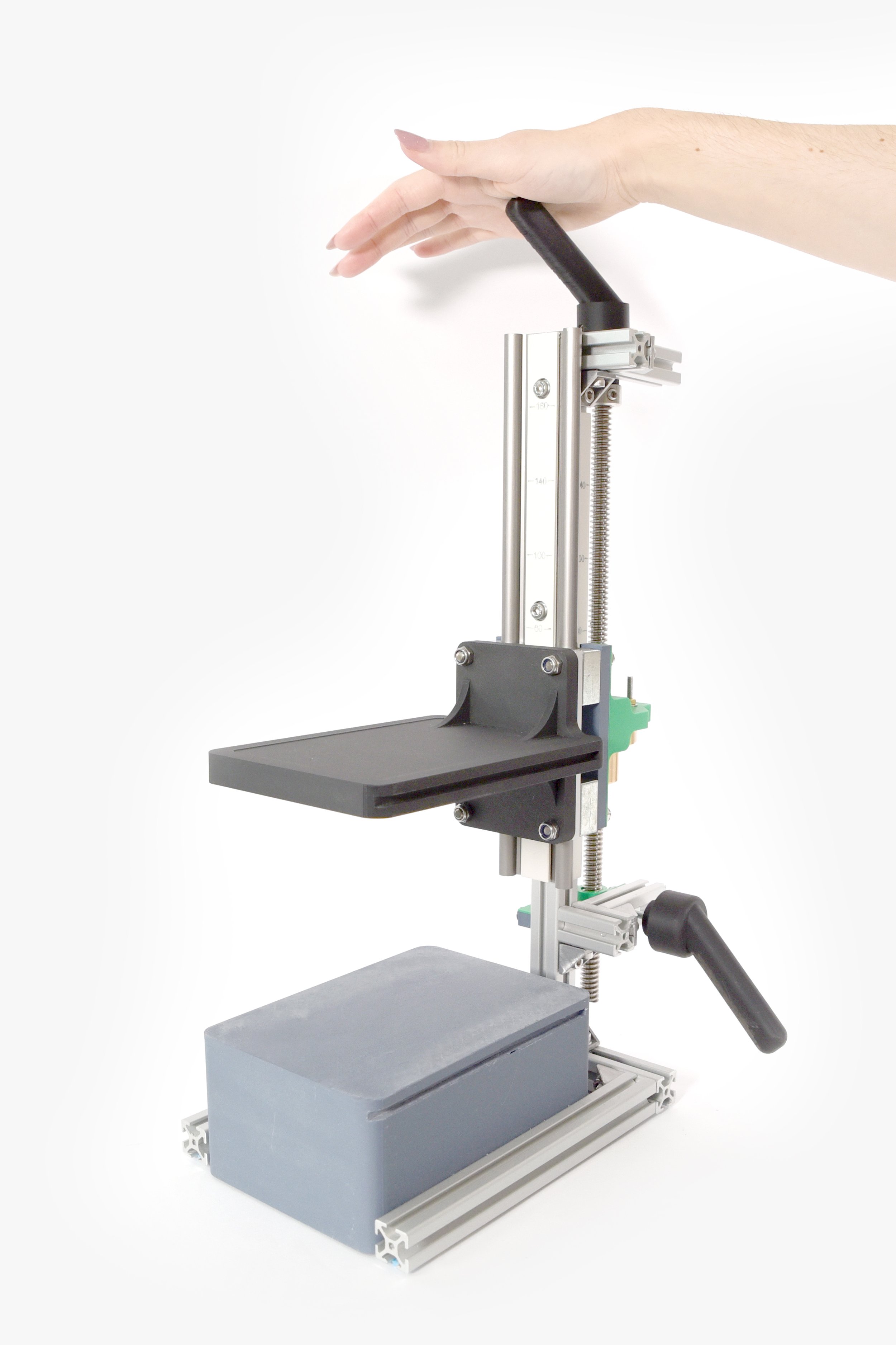

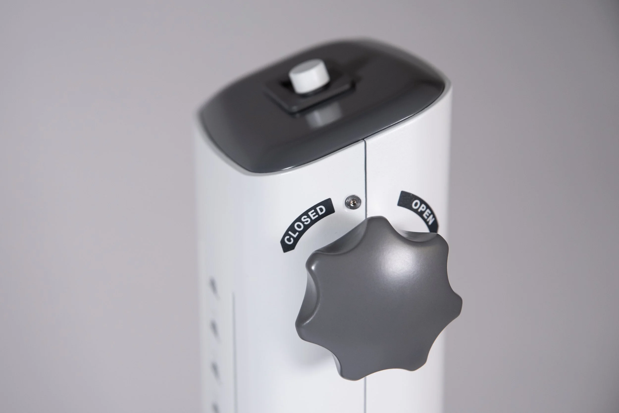
The movement of the sensor assembly was controlled by several sets of dials, geared differently to provide both fine and gross control. This meant researchers could close the platform in a slow and controlled way, but also open it again quickly should there be an issue.
To understand how different parameters affected performance as the device closed around the breast, the relative distance as well as the force it applied were quantified.
For this initial research platform, we used an adapted vernier caliper and shear beam loadcell that would display via an Arduino controller on the top of the device where the researcher could monitor in real-time, and simultaneously output readings to a simple custom datalogger for later analysis.
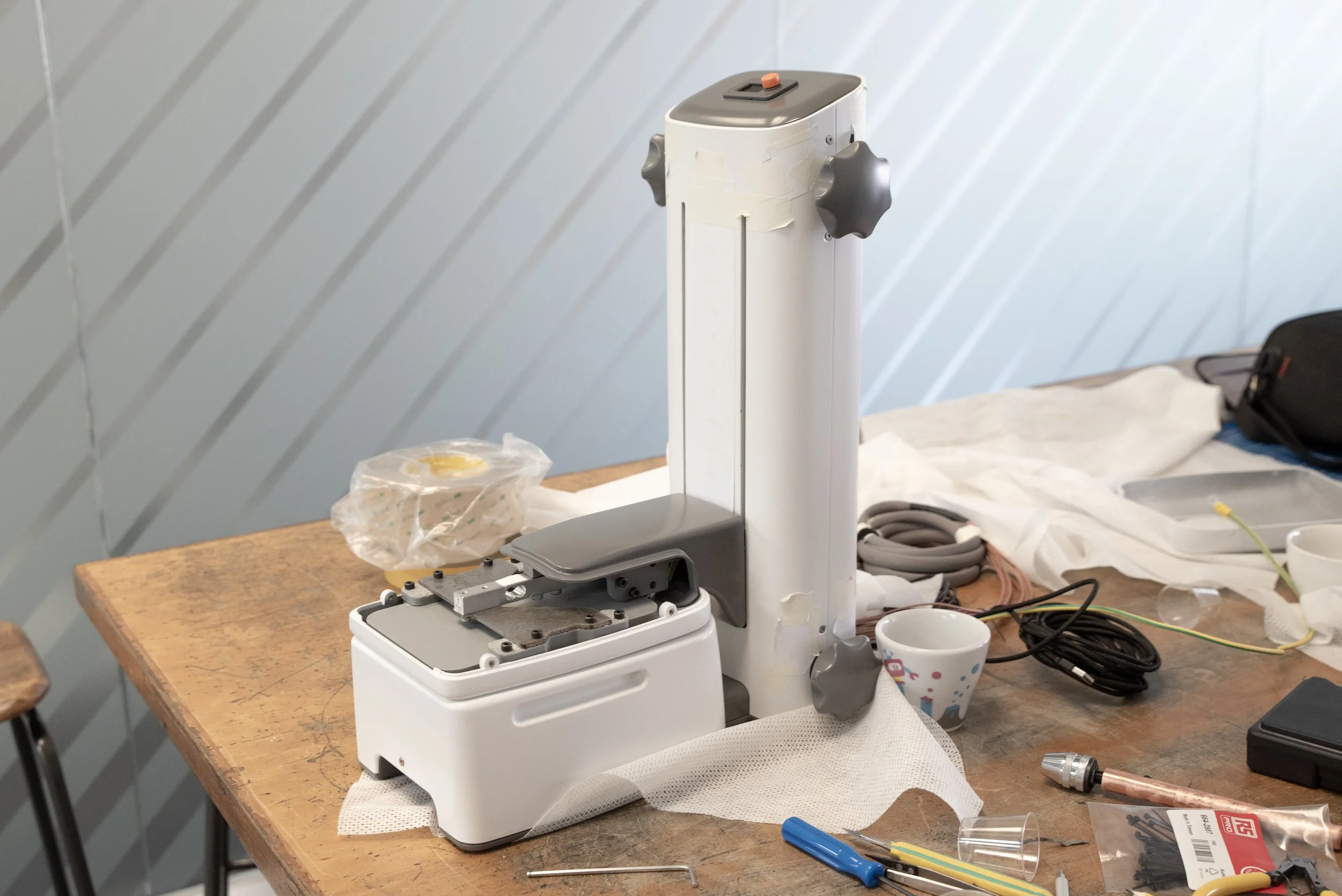
Whilst this is a research platform only, we wanted to address some of the anticipated usability requirements that might exist in practice at point of care.
To accommodate larger participants, the device incorporated accessory wings to support tissue that fell beyond the scanning area which conveniently located and affixed with concealed magnets.
As with more typical ultrasound examination, part of expelling air requires application of a lot of ultrasound gel. Parting lines, fixtures and blind corners were minimised near the scanning plane to facilitate thorough cleaning between participants.
Once NPL have completed their research program, we’re hoping they’ll be able to provide feedback that informs areas for optimisation or development to progress a version of the device towards wider use.